
PLATFORMS TRAINING
The students enrolled in the first year of the Master of Neurosciences have the opportunity to receive advanced training on research platforms in Lyon.
Contact : Emiliano Macaluso and Marion Richard (platforms.neurograduate [at] univ-lyon1.fr)
For the academic year 2024-25, four platforms are available:
- Magneto-encephalography
- Virtual Reality
- From behavior to neurochemistry and vice-versa
- Explore brain function in zebrafish
Magneto-encephalography
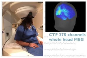
Located in Vinatier Hospital, the MEG department of CERMEP is equipped with a whole head MEG system (CTF – 275 sensors) and numerous associated stimulation devices. The system is used in both basic neuroscience and clinical research in Human by users coming from CRNL, ISCMJ, SBRI, HCLs and other institutions. Two engineers are working full-time in the MEG department to support investigators along the experimental procedure from the protocol definition to advanced data analysis. When activated, large populations of neurons generate a magnetic field of the order of 10-100 fT, one billion times smaller than the earth’s magnetic field (60 microT). Magnetoencephalography (MEG) non invasively measures this magnetic field near the scalp with whole-brain coverage and a temporal resolution of ms, in line with the speed of neuronal processing.
The MEG training program intends to familiarize students with how to design a neuroimaging experiment in human cognitive neuroscience given initial question and hypothesis, and how to perform it, from its implementation through to data analysis and the synthesis of the main results. In particular, we will focus on Magnetoencephalography (MEG) which offers non invasiveness as well as both high temporal and spatial resolution (a technique that is used in fundamental and clinical research, with only 5 such devices in France).
Prerequisites
Students should be highly motivated to learn programming, and we will support them through guidance, online tutorials, and documentation, which may involve some personal investment. They should have knowledge as well as statistics (distributions, t-test/ANOVA, regressions). Here are a some internet resources that would be useful for this: 1) Videos on YouTube; 2) A course with only text and tasks; 3) Some additional resources @intelligent and @bestcolleges.
Main learning outcomes
- formulate hypotheses, design and code an experiment
- record and preprocess behavioural and MEG data
- work with NumPy arrays and Pandas DataFrames at a basic level, and create plots using Matplotlib
- perform time-frequency and statistical analysis on MEG data
- recognize the challenges and strengths of MEG research
Virtual Reality
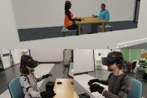
The Neuro-Immersion platform aims to provide researchers with equipment and skills to create experimental devices including virtual reality. To do this, we have virtual reality equipment, such as wired (HTC Vive Pro) and wireless (HTC Vive Focus) VR headsets equipped with eye-trackers, infrared motion tracking systems (VICON), 3D projector, haptic jackets. But also more classic neuroscience systems such as an EEG, a TMS device, a tDCS device, tactile stimulators, biological signal sensors.
During the training, students will be able to create a protocol including technologies available at Neuro-Immersion, this year particularly in the context of multisensory perception during real navigation in a digital virtual space. After an initial presentation by the students, their protocol will be refined in collaboration with the platform team to establish specifications describing the details of the experience. Then, with the support of the platform’s technical team, the students will be able to create the experimental device corresponding to the developed protocol. They will then be able to use this device to acquire experimental data. Finally, students will be able to process and analyse the data acquired using dedicated tools.
From behavior to neurochemistry and vice-versa
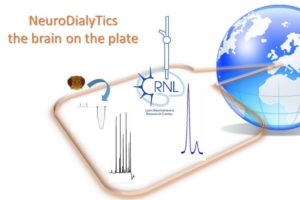
Created in 2014, installed and operated by neurochemists, thanks to its expertise and methodological developments in in vivo and ex vivo neurochemistry, NeuroDialyTics is a facility specialized in the collection, handling and analysis of neuroactive compounds present in very low-volume samples collected for experimental research (rodents, rabbits, flies, crustaceans). The available analytical tools and protocols were customized according to questions initially asked by previous research projects. In such a scientific context, a long-standing collaboration between NeuroDialyTics and WAKING team is at the origin of a unique expertise gained in analyzing and quantifying neurotransmitters in single fly brain samples with microseparative methods which are uncommon and poorly mastered in neuroscience labs (e.g., our use of nano or micro-HPLC).
The proposed training activities rely on very recent developments that permit the determination of up to 18 brain amino acids with a protocol now available on the platform and tested in vertebrate and invertebrate minute brain samples. With the fruit fly Drosophila, the method is sufficiently sensitive to evaluate the content of up to 16 amino acids in a single brain, allowing to link amino acid availability with the feeding and sleep/wake history of an individual fly. Free amino acids levels are modulated by the circadian clock and neuronal activity. It will be asked to the students to make a survey of the scientific literature on the link between metabolism, circadian rhythms and sleep/wake, to propose an original study in accordance with both the literature and the technical framework included in the proposal. As the framework is very open, the subject of the study that will be chosen by the students is rather free and could constitute a valuable pilot study. Indeed, if the results were novel and sufficiently scientifically solid, they could be included in a scientific publication from the WAKING team acknowledging the participation of the students.
Prerequisites
- curiosity and ability to screen through articles to identify unanswered scientific questions
- appetite for cellular and molecular aspects of neurobiology
Main learning outcomes
- learn how to design from scratch an experimental strategy that can address a scientific question
- understand the principles and practices of sample preparation and analysis (here with HPLC)
- perform behavioral analysis of sleep in an animal model
- learn how to analyze data and interpret the results of an experiment, taking into accounts its technical limitations
- discover and experience a full overview of the scientific and technical process in a real project
Biosensors / metabolism in rats
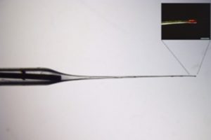
BELIV is an open CRNL technological platform offering in vivo bioelectrochemical techniques. The platform develops neurochemical phenotyping techniques involving implantable microelectrode biosensors to detect and quantify real time concentrations changes of neurochemicals (oxygen, glucose, lactate, glutamate, choline, D-serine) in the brain and other targeted organs. BELIV platform is particularly recognized for developing implantable sensors for brain monitoring. It also offers an in vivo platform for implanting bioelectrochemical devices such as biofuel cells in rats.
As part of the training activities we offer students the opportunity to familiarize themselves with this biosensor technique for assessing metabolic fluctuations induced by different anesthetic agents in rats. The techniques that the students will explore at the BELIV platform allow to measure the real time changes of metabolites (e.g. glucose, lactate) or oxygen, combine with cerebral vascularization using video-microscopy or laser Doppler flowmeters. The effect of different anesthesia conduction is still a matter of debate. This experimental setup allows to investigating different anesthetics used in patients (isoflurane, propofol or thiopental) with light or deep sedation levels. The specific methods and overarching subject matter will be introduced to the students at a preliminary gathering. Following this introduction, students will have the opportunity to explore their individual scientific inquiries within the established context. Examples of such inquiries might include examining the relation between glucose and lactate levels under varying anesthetic conditions; investigating the alterations in cerebral metabolic rate of oxygen (CMRO2) and glucose in response to different anesthetics; and analyzing the connection between metabolic processes, cerebral blood flow, and fluctuations in brain activity. The students will be supported by Dr Baptiste Balança (intensive care anesthetist at the HCL and researcher with the CRNL’s TIGER team) in setting up their project, and by Dr Anne Meiller (engineer with the CRNL’s BELIV platform). Upon approval of the project, students will be trained in the fabrication and application of biosensors. Subsequently, they will employ these biosensors in practical experiments on rats under anesthesia during two brain surgeries performed by Dr. Balança. This hands-on experience will provide them with data that they will compile and examine as the outcome of their research project.
Explore brain function in zebrafish
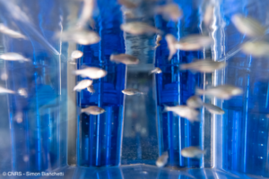
In the framework of the current training activities, the students will use the zebrafish model to tackle an unexplored scientific question on the role of minor splicing in neurodevelopment (project in collaboration with the GENDEV team of the Centre de Recherche en Neurosciences de Lyon). Students will have to design an original scientific project to explore brain function in a zebrafish model of a rare genetic disease, characterized by microcepahly, brain abnormalities and intellectual deficiency. They will have the opportunity to set up crosses of adult fish, collect embryos, observe larvae between 2 and 5 days post-fertilization, conduct behavioral tests, and manipulate them before proceeding to confocal imaging. Part of the work will be carried out in the facility dedicated to zebrafish, and the other part at the CRNL, with members of the GENDEV team.
Prerequisites
None
Main learning outcomes
- design an experiment to address a scientific question
- perform zebrafish experiments
- perform confocal imaging
- analyse images using ImageJ